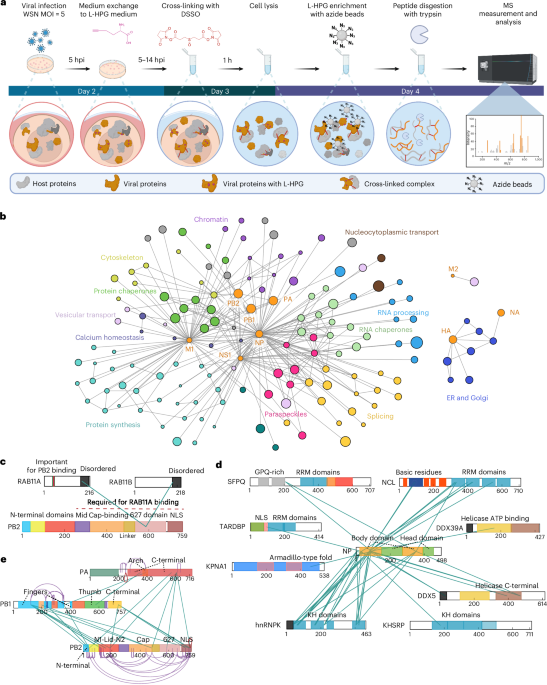
Researchers reporting in Nature used an in-cell protein mapping approach to examine what happens inside human cells during influenza A infection. By applying cross-linking mass spectrometry in infected cells, the team tracked which proteins come into contact, creating a closer view of how the virus reorganizes the host environment to support its own growth.
The study points to two major effects. First, influenza A appears to take over host membrane-trafficking factors, suggesting the virus repurposes normal cellular transport machinery during infection. Second, the work indicates that infection breaks down paraspeckles, a set of structures found in the cell nucleus, and that this disruption helps the virus replicate more efficiently.
Rather than looking at viral components in isolation, the research focuses on protein interactions inside living cells, which can expose changes that are missed by more limited methods. According to the methods snippet, the experiments used immortalized human cell lines including A549 and HEK293T, giving the researchers a controlled system for comparing infected and uninfected cells.
Taken together, the findings add to the picture of influenza A as a virus that does more than enter a cell and copy itself. It actively reshapes protein networks, redirects host machinery and alters nuclear structures in ways that appear to favor viral replication.